
The worldwide increase in cancer cases is a substantial impediment to extending how long people live. Over the last few decades, extensive research has been conducted to find ways to conquer this lethal disease. As a result, cancer treatments such as the three major cancer treatments, molecular targeted therapy, and immunotherapy have made significant progress. However, these methods still have limitations, such as severe side effects, invasiveness, and an inability to effectively treat terminal stages.
In this blog, I would like to inform you of the study, “Real-Time Monitoring of Colorectal Cancer Location and Lymph Node Metastasis and Photodynamic Therapy Using Fucoidan-Based Therapeutic Nanogel and Near-Infrared Fluorescence Diagnostic–Therapy System” by Yoo-kyoung Shin et al. The study reported on real-time monitoring of colon cancer, lymph node metastasis of colon cancer cells, and tumor growth inhibition by photodynamic therapy (PDT) using a near-infrared fluorescence diagnostic and treatment system equipped with a PDT light source and a fucoidan-based theranostic nano gel (CFN gel) with excellent accumulation efficiency in cancer cells.
In the study, researchers report on the possibility of image-guided surgery of colon cancer by PDT using the CFN-gel and near-infrared fluorescence diagnostic and treatment system they have developed, as well as the possibility of suppressing lymph node metastasis and tumor growth of colon cancer cells. First, they performed the diagnosis of colon cancer and confirmation of metastasis by fluorescence imaging using three laser diodes with the absorption wavelengths and treatment wavelengths of PpIX (5-ALA), Ce6, and CFN-gel used in this study, 405 nm, 650 nm, and 660 nm. Additionally, an optical switch was utilized to establish a near-infrared fluorescence diagnostic and treatment system.
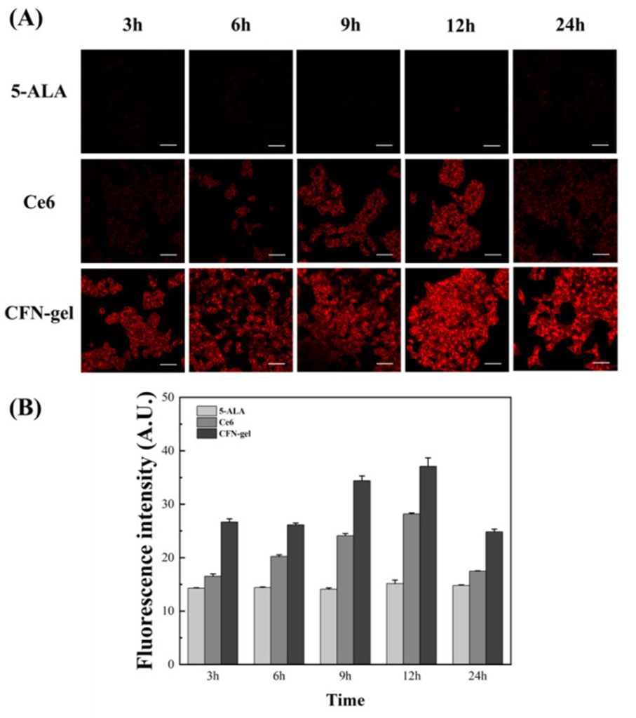
In order to validate the accumulation of the contrast agent injected into HCT 116 cancer cells more accurately and compare the resulting images with a confocal microscope, we employed a custom-made two-photon microscope to capture images of 5-ALA, Ce6, and CFN-gel, as shown in Figures 1 and 2. In the case of CFN-gel, the binding to colon cancer cells initially showed low fluorescence intensity. However, it increased significantly after 6 hours, and even after 24 hours, the fluorescence intensity was higher than that of 5-ALA and Ce6.
By utilizing both one-photon imaging and two-photon imaging, the accumulation and fluorescence intensity of PpIX (5-ALA), Ce6, and CFN-gel in cancer cells can be verified. In the case of CFN-gel, the fluorescence intensity was maintained even after 24 hours. In this way, the binding and fluorescence intensity of PpIX (5-ALA), Ce6, and CFN-gel in cancer cells could be confirmed by one-photon imaging and two-photon imaging. CFN-gel’s ability to maintain fluorescence intensity for over 24 hours makes it an ideal solution for accurately identifying cancer location in real-time during colorectal surgery.
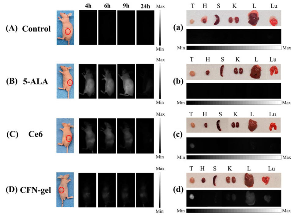
The possibility of image-guided surgery using the proposed imaging system and the previously developed CFN-gel was confirmed through in vivo experiments on xenograft mice injected with colon cancer cells, as shown in Figure 2. The accumulation of cancer cells was observed over time by injecting commercially available 5-ALA, Ce6, and CFN-gel. The findings indicated that 5-ALA does not accumulate significantly at the tumor site but is eliminated from the body over time. Figure 1B shows that mice administered 5-ALA emitted strong fluorescence throughout the whole body after 4, 6, and 9 hours.
Similar to the results of the cancer cell experiment, it was found that Ce6 was more highly accumulated than 5-ALA in the cancer area of xenograft mice. The necessity to establish whether PpIX (5-ALA), Ce6, and CFN-gel had accumulated in the mouse body posed a challenge. Consequently, a decision was made to sacrifice each mouse after 24 hours to perform a thorough examination of the tumor and organs, including the heart, spleen, kidney, liver, and lung, for fluorescence. No fluorescent signal was observed in the tumor and organs for 5-ALA, and it was found that most of PpIX (5-ALA), Ce6, and CFN-gel had been excreted from the body after 24 hours. However, in the case of Ce6, a fluorescent signal was observed in the tumor, although the fluorescence intensity was low. CFN-gel not only showed high fluorescence intensity in the tumor but also a small amount of fluorescent signal was confirmed in the lung and liver. Thus, CFN-gel was well adsorbed to colon cancer and had a long circulation time in the blood, confirming the feasibility of image-guided surgery using the proposed system.
The proposed fluorescence imaging system and the previously developed CFN-gel were used to confirm the possibility of diagnosing and treating colon cancer. Lymph node metastasis was confirmed by fluorescence imaging and verified by H&E staining. Clear fluorescent images were confirmed in the lymph nodes of a mouse model using CFN-gel. Although cancer cells were present throughout the lymph nodes, no fluorescence from PpIX(5-ALA) or Ce6 was observed with H&E staining, and CFN-gel enabled confirmation of lymph node metastasis in real-time with clear fluorescent images. This finding demonstrates that the combination of CFN-gel with the proposed near-infrared fluorescence diagnostic and treatment system allows for the simultaneous diagnosis and treatment of colon cancer and lymph node metastasis.
The study has demonstrated the viability of image-guided surgery and PDT when the proposed system is combined with CFN-gel. Hence, the conclusions drawn from this research may lay the groundwork for advanced image-guided surgery in the future, and could potentially become an essential technology for immediate confirmation of metastasis in the lymph nodes adjacent to the surgical site.
Source: Pharmaceutics. 2023 Mar; 15(3): 930. doi: 10.3390/pharmaceutics15030930