
Osteosarcoma primarily impacts adolescents and is a form of bone cancer. The negative outcomes of chemotherapy, surgical complications, and the potential for postoperative osteoporosis all contribute to a poor prognosis and diminished quality of life following treatment. Fucoidan, a type of sulfated polysaccharide found in brown algae, has gained significant interest due to its potential anti-cancer effects and its ability to promote bone regeneration.
In this blog, I would like to share the study, “The Effects of Fucoidan Derived from Sargassum filipendula and Fucus vesiculosus on the Survival and Mineralisation of Osteogenic Progenitors” by Dhanak Gupta et al. The study concentrated on human embryo-derived mesenchymal stem cells, the researchers investigated the effects of crude low molecular weight (LMW, 10-50 kDa), medium molecular weight (MMW, 50-100 kDa), and high molecular weight (HMW, >100 kDa) fractions from Sargassum as well as crude fucoidan from Fucus vesiculosus.
First, in FTIR-ATR analysis, all fucoidans showed characteristic absorption bands of sulfated polysaccharides, respectively, indicating that the crude extract of F. vesiculosus had significantly higher sulfation levels and glycosidic bonds and lower uronic acid content compared to all other fucoidans.
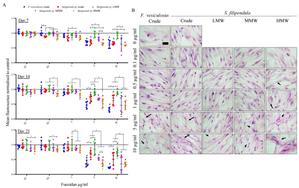
The study findings revealed a consistent and dose-dependent decrease in metabolic activity of hES-MP cells when exposed to different types of fucoidan, indicating a notable impact on cell proliferation and morphology. Notably, the most prominent effect was observed at a concentration of 10 μg/mL of F. vesiculosus fucoidan, as illustrated in Figure 1A.
Furthermore, fucoidan derived from S. filipendula showed a molecular weight-dependent effect on cellular metabolic activity. Cells treated with the LMW fraction showed the highest metabolic activity, followed by the MMW fraction, and the lowest metabolic activity in cells treated with the HMW fraction, at all time points, especially at doses above 1 μg/mL. Analysis of Giemsa staining performed on day 21 (See Figure 1B) revealed that cells in the control condition displayed a distinct elongated and flattened shape, accompanied by nuclei that were well-defined. For HMW, this adverse effect on cell morphology was also seen at low doses of 0.5 and 1 μg/mL, whereas for the other Fucoidans, toxic doses were higher than 1 μg/mL. The obtained results provide evidence that fucoidan has the ability to suppress cell growth and disrupt the cytoskeleton in a way that varies according to the dosage, with the HMW fraction having the most detrimental effect.
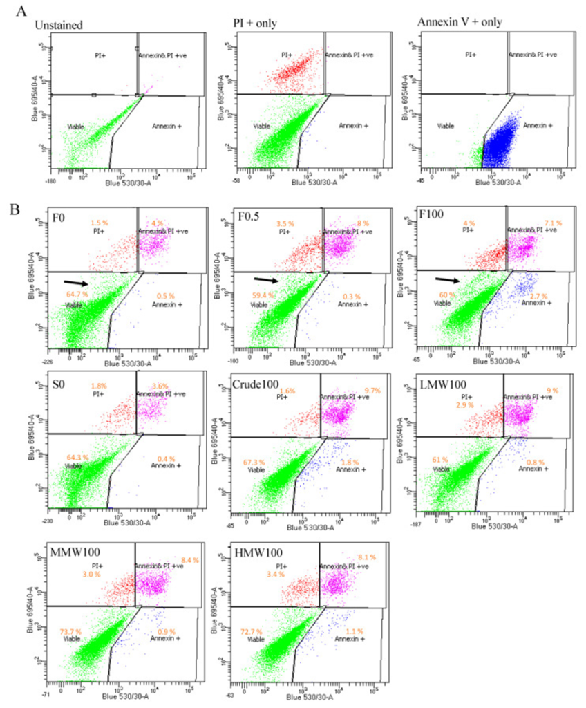
Annexin V/propionium iodide (PI) staining was performed. The results, as depicted in Figure 2, clearly demonstrated that the presence of F. vesiculosus fucoidan at concentrations of 0.5 and 100 μg/mL led to a significant decrease in viable cells and a notable increase in both early apoptotic and dead cells when compared to the respective solvent control with no F. vesiculosus fucoidan (0 μg/mL). To further confirm the observations, cells were seeded at a higher cell density and treated with F. vesiculosus-derived fucoidan at 0, 0.5, and 100 μg/mL for 10 days. The findings indicated that crude fucoidan from F. vesiculosus resulted in lower cell viability, increased apoptosis, and a noticeable accumulation of unstained cell debris.
Additionally, the results revealed a significant increase in the early apoptotic cell population following treatment with F. vesiculosus crude fucoidan, in contrast to S. filipendula-derived crude fucoidan. Furthermore, there was also an increase in late apoptotic cells between fractions as MW increased. These results indicate that fucoidan derived from F. vesiculosus induced apoptosis-like cell death, whereas fucoidan derived from S. filipendula led to cell necrosis, especially in the high molecular weight fraction.
To investigate whether mitochondrial integrity is affected by fucoidan, Transmission electron microscopy (TEM), JC-1 (5,50,6,60-tetrachloro-1,10,3,30-tetraethylbenzimidazolylcarbocyanine iodide) staining, and cytochrome c analysis confirmed fucoidan-induced mitochondrial damage, ER swelling, and enhanced autophagy, which was most severe with F. vesiculosus fucoidan. They also observed stress-induced cell death similar to apoptosis with F. vesiculosus fucoidan, and cell death resembling necrosis with S. filipendula fucoidan. LMW and MMW doses below 200 ng/mL were the least toxic and showed potential osteoinductivity. The study sheds light on the various effects of fucoidan on osteoblasts and underscores the delicate trade-off between potential therapeutic benefits and challenges when using fucoidan as a postoperative treatment for osteosarcoma patients.
Also, the results from studies suggest that delivery of low doses of fucoidan in a controlled manner over an extended period to the bone regeneration site containing stem cells after osteosarcoma resection (possibly in combination with a 3D scaffold) could promote bone formation in that area and damage remaining cancer cells. Based on the current results, the most potent fucoidans were the LMW and MMW fractions of S. filipendula (in that order) in the 10-100 kDa range. However, additional studies are required to identify the specific molecular weight ranges that can effectively achieve dual therapeutic objectives.
Source: Int J Mol Sci. 2024 Feb 8;25(4):2085. doi: 10.3390/ijms25042085