The administration of cytostatic agents to chemotherapy in cancer patients has significant side effects, including inhibiting various parts of hematopoiesis.
Hence, in this blog, I would like to introduce a study, “Fucoidan and Fucosylated Chondroitin Sulfate Stimulate Hematopoiesis in Cyclophosphamide Induced Mice” by Natalia Anisimova et al. The following study showed the results of fucoidan for the concomitant treatment of cancer patients who suffered hematologic toxicity during chemotherapy and radiotherapy.
This study also indicated two sulfated polysaccharides as stimulators of hematopoiesis, fucoidan (PS-Fuc) from the seaweed Chordaria flagellum and fucosylated chondroitin sulfate (PS-FCS) from the sea cucumber, Massinium magnum were associated with hematopoiesis after cyclophosphamide immunosuppression in mice, and recombinant granulocyte colony-stimulating factor (rG-CSF) was applied as a reference.
First, thirty male mice of the CBA strain were divided into five groups of 6 mice each. Cyclophosphamide (CPh) was injected intraperitoneally to induce myelosuppression at a dose of 100 mg/kg into four groups of animals once daily for four days. They administered the following sterile solution (0.2 mL) subcutaneously to all animals for three days (once daily). All animals were given the following pure solutions (0.2 mL) subcutaneously for three days (once daily), also administrated isotonic sodium chloride solution of PS-FCS (group CPh + PS-FCS), and isotonic sodium chloride solution (group CPh + r G-CSF), sterile isotonic sodium chloride solution (group CPh).
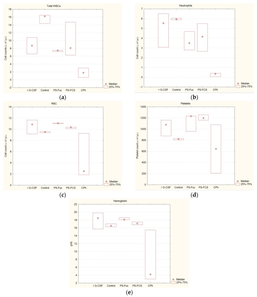
A clean isotonic sodium chloride solution was administered to a control group of mice with the same regimen. The results of hematological parameters in different groups of mice are shown in Figure 1. White blood cells (WBC), neutrophils, Red blood cells (RBC), platelets, and hemoglobin levels were measured. They significantly increased WBC compared to group CPh under the influence of PS-Fuc, PS-FCS, and rG-CSF. It indicated a normalization of CPh-induced leukopenia after administering PS-Fuc and PS-FCS.
At the same time, their activity levels were not significantly different from those of rG-CSF. Restoration of total WBC counts under the influence of both test compounds was due to increased neutrophil counts in mice with CPh-induced leukopenia. This trend was more evident under the influence of PS-FCS and rG-CSF than under the influence of PS-Fuc. Neutrophil counts increased 10.5-, 13.8-, and 8.8-fold, respectively, compared to the CPh group.
After treatment with rG-CSF, PS-Fuc, and PS-FCS, we observed increased RBC, hemoglobin, and platelets in mice immunosuppressed by CPh. Increases in RBC, hemoglobin, and platelets were statistically significant compared with controls when PS-Fuc and PS-FCS were used. In contrast, the effect with rG-CSF was not substantial as p-values significantly exceeded 0.05. (0.226 for RBC, 0.328 for hemoglobin, and 0.063 for platelets)
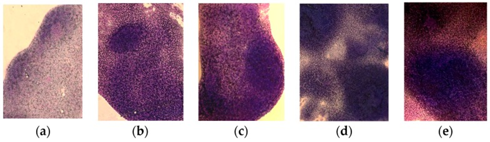
Analysis of splenic morphology on smears showed that myelosuppression was accompanied by depletion of the cellular composition of the white pulp, represented mainly by WBCs, after repeated administration of CPh. (Figure 2a) Thus, follicles (B-dependent regions) and peri-arteriolar sheaths (T-dependent regions) disappeared. The splenic architecture was disrupted, and the interstitial tissue consisted of a dense, uniform layer of lymphoid cells.
Therefore, both PS-Fuc and PS-FCS polysaccharides were tested, and cell repair was observed after rG-CSF (Fig. 2b–d). Especially an intensive recovery was seen after the PS-FCS administration.
As a result, these compounds can be viewed as potentially promising hematopoietic stimulators. Furthermore, this study proves that such drugs will be in demand for the concomitant treatment of cancer patients who suffer hematologic toxicity during chemotherapy and radiotherapy.
Source: Mar Drugs. 2017 Oct; 15(10): 301. doi: 10.3390/md15100301