
Doxorubicin (DOXO), a chemotherapy medication, treats various cancers, including breast cancer, solid tumors, leukemia, and soft tissue sarcoma. However, the therapeutic value and clinical application of DOXO are limited by both acute and chronic life-threatening cardiotoxicity, including arrhythmias, tachycardia, and even severe congestive heart failure and left ventricular dysfunction. Doxorubicin causes cellular atrophy; its induction increases cardiac volume and causes hypertrophy.
Fucoidan, on the other hand, is a naturally occurring, active sulfate polysaccharide that has been shown in various studies to possess a wide array of biological activities, including anticoagulant and antithrombotic effects, antitumor properties, antiviral and antioxidant capabilities, and anti-inflammatory actions. Recent studies show fucoidan’s strong antioxidant effects both in the lab and in living organisms. Furthermore, fucoidan is involved in improving mitochondrial function and alleviating oxidative stress.
In this blog, I would like to share the following study, “Fucoidan Protects against Doxorubicin-Induced Cardiotoxicity by Reducing Oxidative Stress and Preventing Mitochondrial Function Injury” by Yuting Ji et al. The research indicated that oral fucoidan supplementation exerts a strong protective effect against DOXO-induced cardiotoxicity by reducing oxidative stress and preventing mitochondrial dysfunction.
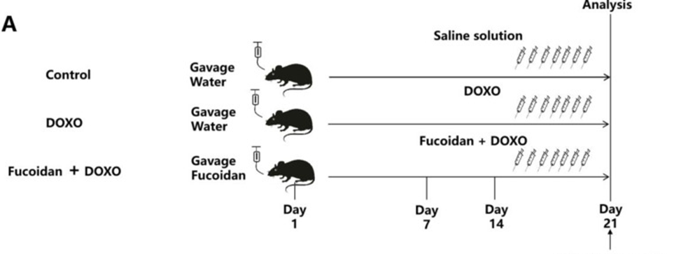
The first experiment involved investigating the protective effects of fucoidan against the cardiac injury induced by DOXO in mice, and the results of this experiment are presented in Figure 1A. Group 1 mice were given only water for 21 days (control group, CTL). Group 2 mice were given water for 14 days and then intraperitoneally injected with 7 mg/kg DOXO daily for 7 days (DOXO group). Group 3 mice were given fucoidan for 14 days first, then DOXO and fucoidan together for 7 days (fucoidan + DOXO group).
In vivo transthoracic echocardiography was used to investigate the cardioprotective effects of fucoidan against DOXO-induced toxicity by examining several indicators. The results showed that the left ventricular function of mice was significantly damaged after the DOXO injection compared with the CTL group. However, DOXO injection-induced cardiac injury could be improved by oral administration of fucoidan. In the mice detection of the fucoidan + DOXO group, both returned to the level of the CTL group, suggesting the protective effect of fucoidan.
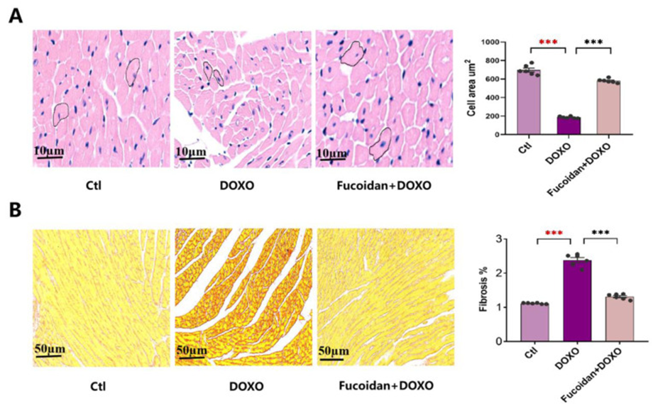
To investigate whether fucoidan could attenuate DOXO-induced myocardial atrophy and cardiac fibrosis, two important markers of DOXO-induced cardiotoxicity, cardiomyocyte size, and cardiac fibrosis were analyzed by H&E staining and picrosirius red staining. Cardiomyocytes in the DOXO group were smaller and exhibited increased interstitial space compared to the CTL group, as revealed by H&E staining. However, these lesions were significantly improved in mice orally administered fucoidan (See Figure 2A). In addition to previous findings, picrosirius red staining revealed a notable increase in interstitial fibrosis following DOXO injection, an effect that fucoidan treatment significantly mitigated, as evidenced by the results depicted in Figure 2B.
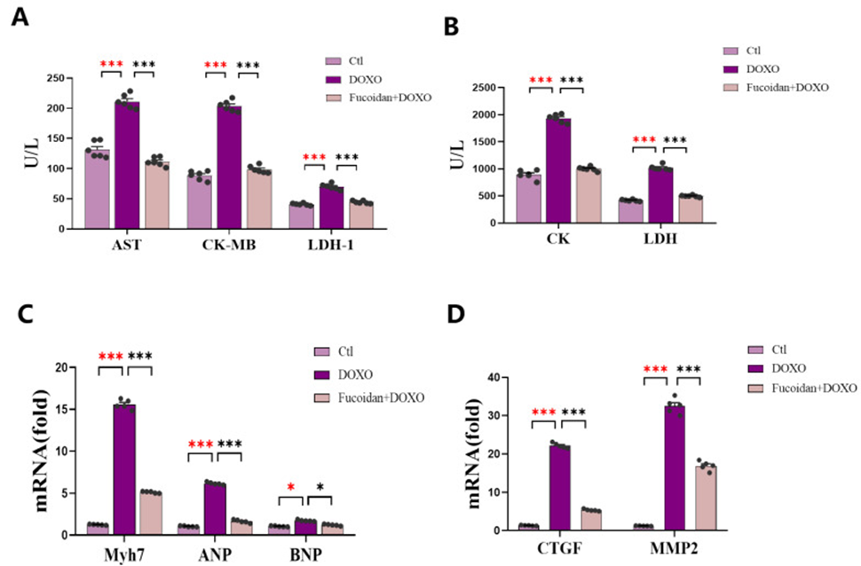
Measuring the serum levels of enzymes characteristic of cardiac function, the contents of AST, CK and its isozyme CK-MB, and LDH and its isozyme LDH-1 in mice in the DOXO group were significantly higher than those in the CTL group (See Fig. 3A, B). In contrast, fucoidan was able to reduce the expression levels of these factors, which suggests a potential mechanism by which fucoidan might alleviate the myocardial damage caused by DOXO, as illustrated in Figures 3A and 3B. The three groups also showed detectable mRNA expression levels for genes associated with cardiac dysfunction.
The mRNA expression of atrial natriuretic peptide (ANP), brain natriuretic peptide (BNP), and myosin heavy chain 7 (Myh7) was increased in mice in the DOXO group, whereas the expression of these genes was decreased in mice pretreated with fucoidan compared with mice in the DOXO group (See Fig. 3C). Meanwhile, the expression of connective tissue growth factor (CTGF) and matrix metalloproteinase-2 (MMP-2) was significantly increased after DOXO injection, which was consistent with the increase in ANP and BNP, and co-treatment with fucoidan could attenuate these increases (See Fig. 3D). The results demonstrated that administering fucoidan orally protects the heart.
High levels of ROS in cells are a key sign of oxidative stress; all mouse heart tissue samples showed elevated ROS, but fucoidan treatment improved this. Similarly, the levels of MDA (the most common by-product of lipid peroxidation) in the serum of mice indicated that DOXO increased the risk of lipid peroxidation and this process was inhibited by fucoidan. Concurrent with serum measurements of the antioxidant enzyme activities GSH-PX and SOD, a significant reduction in the activity of these enzymes was observed in mice receiving DOXO injections; however, the administration of fucoidan mitigated this reduction and consequently ameliorated the resulting oxidative damage.
Due to the critical role mitochondria play in maintaining the viability of cardiomyocytes, the detection of mitochondrial dysfunction is essential for diagnosing DOXO-induced cardiotoxicity. They detected a significant increase in ROS in DOXO-induced myocardial injury. They then examined the ATP levels in mouse cardiac tissue from all three groups, which showed that the ATP levels were lower in the DOXO group than in the CTL group, but were restored in the fucoidan + DOXO group.
Mitochondrial ATP production is strongly linked to the gene expression of the mitochondrial electron transport chain. Therefore, they examined the expression of mitochondrial cytochrome b (mt-Cytb) and mitochondrial ATPase6 (mt-ATP6), which are encoded in mitochondrial DNA. The mRNA expression levels of these two genes were decreased in the DOXO group but were restored in the fucoidan + DOXO group. In addition, they measured the mRNA expression of marker genes related to the mitochondrial respiratory chain complex І–V in the cardiac tissue of each mouse. The lower gene expression levels in the DOXO-treated group versus the control group indicate that DOXO likely inhibits mitochondrial respiratory chain complexes, thus lowering ATP production. Fucoidan has been shown to have a significant positive impact on improving the efficiency of these processes. The findings of the study indicate that the deleterious effects of DOXO on the functionality of cardiac mitochondria can be mitigated through the administration of fucoidan.
Source: Int J Mol Sci. 2022 Sep; 23(18): 10685. doi: 10.3390/ijms231810685