In this article, I would like to share how fucoidan prevents M2 macrophage differentiation and also discuss how fucoidan differentiates M2 through a study.
Macrophage infiltration within and around the tumor nest is one of the essential hallmarks during tumor progression. According to Jiuyang Liu et al., “New insights into M1/M2 macrophages: key modulators in cancer progression,” Mutual interactions with tumor cells and the stromal microenvironment contribute to the phenotypic polarization of tumor-associated macrophages.
Macrophages are composed of at least two subgroups, M1 and M2. M1 phenotype macrophages are tumor resistant due to their intrinsic phagocytosis and enhanced anti-tumor inflammatory response. In contrast, M2 possesses a repertoire of tumor-promoting functions, including immunosuppression, angiogenesis, angiogenesis, stromal activation, and remodeling. In addition, the functional features of M2 incorporate a location-related, interconnected, cascade-like response that accelerates the pace of tumor aggressiveness and metastasis.
Hence, I would like to introduce the following study,” Fucoidan Prevents M2 Macrophage Differentiation and Colon cancer Tumor Progression,” by Li-Mei Chen et al., investigating how fucoidan acts on cancer M2 macrophages.
Reactive oxygen species (ROS) generated during intracellular metabolism or triggered by exogenous factors can promote neoplastic transformation and the malignant microenvironment that mediates tumor development.
Oligofucoidan is a sulfated polysaccharide isolated from wakame seaweed. Using human THP-1 monocytes and murine Raw264.7 macrophages, human HCT116 colorectal cancer cells, primary C6P2-L1 colorectal cancer cells, and human MDA-MB231 breast cancer cells, Oligofucoidan on inhibition of M2 macrophage differentiation investigated the effect.
First, Li-Mei Chen and others compared the antioxidant capacity of Oligofucoidan (LMF) (0.5–0.8 kDa) from Sargassum hemiphyllum and high molecular weight fucoidan (HMF) (20–200 kDa) from Fucus vesiculosus. Thus, both LMF and HMF suppressed mitochondrial superoxide levels (Supplementary Figure- S3A).
Likewise, phosphorylation of the NADPH oxidase subunit p47phox on Ser345 was dramatically reduced by LMF or HMF treatment (Supplementary Figure- S3B). Interestingly, THP-1 monocyte-differentiated M2 macrophages treated with LMF or HMF also suppressed the population of M2 macrophages compared with MOCK and isotype IgG controls (Supplementary Figure- S3CD).
HCT116 cells were then treated with DPI (diphenyleneiodonium) and Oligofucoidan (400 g/mL), and pretreated cancer cells were placed in Boyden chamber transwells’ upper compartment co-cultured with THP-1 monocytes and cultivated. 48 hours bottom chamber.
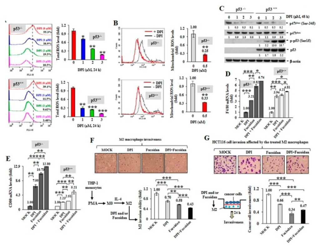
The results showed that DPI-pretreated cancer cells stimulated monocyte polarization towards F4/80 (high) M0 (Figure- 1D) and CD80 (high) M1 (Figure 1E) macrophage phenotypes.
Interestingly, cancer cells treated with Oligofucoidan also upregulated F4/80 and CD80 mRNA levels (Figure- 1D, E), and DPI co-treatment additively enhanced these effects. Therefore, ROS inhibition in cancer cells may indirectly promote M0 and M1 differentiation.
They investigated whether ROS produced in cancer cells affects macrophage plasticity; two isogenic HCT116 colons with no (p53−/−) or (p53+/+) wild-type p53 cancer cell lines were treated with DPI. When DPI suppressed intracellular ROS (Figure- 1A) and mitochondrial superoxide levels (Figure- 1B), phosphorylation of p47phox (Ser345) was significantly reduced by DPI (Fig. 1C), and DPI decreased p53 (Ser15), dramatically induced dynamic phosphorylation.
M2 macrophages were highly invasive (Figure- 1F), but combined treatment with DPI and Oligofucoidan suppressed their invasive potential to a greater extent than the individual treatments. Reciprocally, the invasiveness of HCT116 cancer cells was markedly suppressed when they encountered M2 macrophages pretreated with DPI and Oligofucoidan (Figure- 1G).
Hence, it is observed that DPI and Oligofucoidan treatment can suppress M2 macrophage infiltration and restore M0 and M1 plasticity, preventing cancer cell invasion.
Significantly, Oligofucoidan suppresses the drawback of chemotherapy on M2 macrophage polarization in vitro and inhibits M2 macrophage infiltration in vivo. The results demonstrate that Oligo-Fucoidan supplementation sufficiently enhances chemo-sensitivity in aggressive cancer cells and renovates a healthy microenvironment that prevents tumor progression.

Source: 2020 Feb; 12(2): 421. doi: 10.3390/cancers12020421

Hello my name is Ann my father have liver cancer he’s very ill I would like to no the best fucoidan y’all have to help him thanku
Hi! Ann
I recommend checking the NPO Research Institute of Fucoidan first And you can find the best fucoidan for your father.