Fucoidan, which is the primary constituent of wakame, has been suggested to possess numerous mechanisms that can combat cancer. These mechanisms include the ability to induce cell cycle arrest, promote apoptosis, inhibit angiogenesis, and activate the immune system. Despite evidence of fucoidan’s impact on different HCC cells in both controlled laboratory experiments and living organisms, the molecular events that drive these effects are still not clearly elucidated. As a result, this blog aims to discuss the findings of a study conducted by Ming-De Yan et al, titled “Fucoidan Elevates MicroRNA-29b to Regulate DNMT3B-MTSS1 Axis and Inhibit EMT in Human Hepatocellular Carcinoma Cells”. The study demonstrates the ability of fucoidan to regulate miR expression, leading to the activation of tumor suppressor cascades in HCC cells.
In most human cancers, the expression of miR-29b, the most highly expressed member of the miR-29 family (miR-29a/b/c), is commonly altered. In HCC, decreased expression of miR-29b was observed in clinical samples and was significantly associated with worse disease-free survival of patients, and miR-29b directly targets matrix metalloproteinase 2 (MMP-2) expression. Animal studies have shown that it can effectively block the development of HCC by inhibiting angiogenesis, invasion, and metastasis. According to these studies, fucoidan was found to induce miR-29b and suppress MMP-2 in her HCC cells.
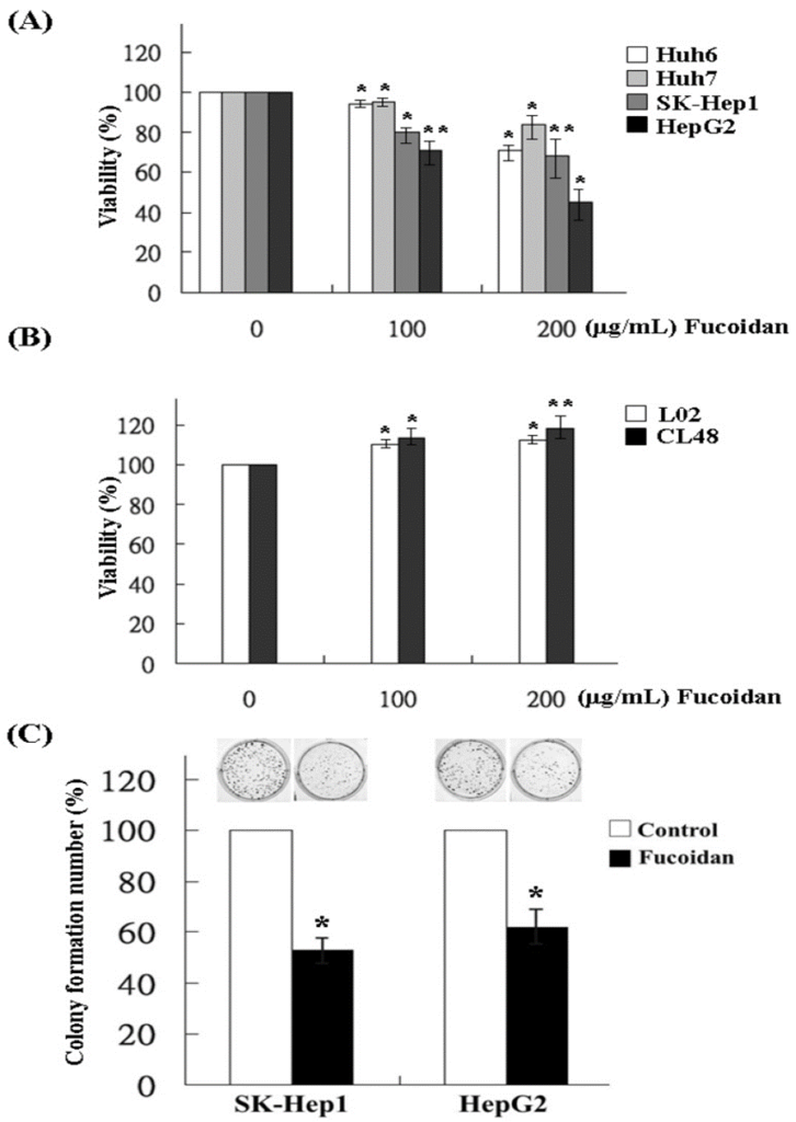
First, the effect of fucoidan on the proliferation of HCC cells was determined by MTS assay in Huh6, Huh7, SK-Hep1, and HepG2 cells. The administration of fucoidan to HCC cells at a concentration of 200 μg/mL for a duration of 48 hours led to the suppression of cell proliferation in Huh6, Huh7, SK-Hep1, and HepG2 cells, as shown in Figure 1A. Unlike normal human liver cell lines, such as L02 and CL48, fucoidan did not exhibit any inhibitory effect on their proliferation. This indicates that fucoidan selectively targets and suppresses the growth of cancer cells, as depicted in Figure 1B. Next, the researchers examined how fucoidan affected the ability of SK-Hep1 and HepG2 cells to form colonies, as these two HCC cell lines were found to be more susceptible to fucoidan compared to the other two cell lines. According to the results, it was observed that at a concentration of 200 μg/mL, fucoidan exhibited a significant reduction in the colony formation of both SK-Hep1 cells and HepG2 cells, as shown in Figure 1C.
Besides the growth of tumors, the invasion and distant metastasis also have a significant impact on the unfavorable clinical outcome of HCC. Hence, a more detailed investigation was conducted to assess the influence of fucoidan on the invasion potential of SK-Hep1 and HepG2 cells, employing the transwell invasion assay. As shown in Figure 2, at a dose of 200 μg/mL, fucoidan significantly reduced the invasion of SK-Hep1 and HepG2 cells. The miR expression profile of Fucoidan-treated HepG2 cells was analyzed by Affymetrix GeneChip miRNA 2.0 Array. The findings from the study indicated that the administration of fucoidan resulted in an increase in the expression levels of tumor suppressor miRs, specifically the miR-29 family and miR-1224. However, fucoidan significantly suppressed the expression of oncogenic miRs, such as the miR-17/92 cluster, in contrast to other miRs.
Prediction of miR-29b targets using Pic Tar and TargetScan databases showed that DNMT3B had the highest score and was considered to be a potential miR-29b target gene. In order to confirm the inhibition of DNMT3B by miR-29b in HCC cells, the researchers created luciferase reporter constructs. These constructs contained the 300 bp 3′-UTR of DNMT3B, with separate versions for wild-type miR-29b binding and mutant miR-29b binding. Wild-type luciferase activity was suppressed to approximately 50% in SK-Hep1 and HepG2 cells after treatment with fucoidan for 48 hours (Figure 3B). This showed that fucoidan increased miR-29b expression and negatively regulated her DNMT3B in HCC cells.
In order to validate the inhibitory effect of fucoidan on DNMT3B, several experimental techniques were employed including Western blot and RT-PCR. The results showed a dose-dependent suppression of both mRNA and protein levels of DNMT3B in fucoidan-treated SK-Hep1 and HepG2 cells. Fucoidan dose-dependently increased MTSS1 mRNA and protein levels while suppressing DNMT3B in these HCC cells. In the following step, the EMT markers in the HCC cells were examined, particularly focusing on the increased expression of MTSS1. Our observations revealed that the upregulation of E-cadherin and the reciprocal downregulation of N-cadherin signify the inhibition of EMT in the fucoidan-treated HCC cells.
Fucoidan also inhibited TGF-β signaling in these HCC cells. Following that, they proceeded to conduct a more extensive investigation into the influences on MMP and TIMP. Fucoidan dose-dependently decreased the protein levels of MMP2 and MMP9, while increasing the protein level of tissue metalloproteinase-1 (TIMP-1). These effects may also contribute to the decrease in the invasive activity of HCC cells shown in Figure 2.
Fucoidan not only inhibits TGF-β signaling but also has a profound effect on the miR-29b-DNMT3B-MTSS1 axis. The observed effects include the inhibition of EMT, characterized by an increase in E-cadherin expression and a decrease in N-cadherin expression. Additionally, they contribute to the prevention of extracellular matrix degradation, as indicated by an increase in TIMP-1 levels and a decrease in MMP2 and MMP9 levels. The combined impact of these effects is crucial in reducing the movement and infiltration of HCC cells.
Based on the results obtained in this study, fucoidan shows promise as an integrative therapeutic agent for improving treatment outcomes in patients with HCC.


Source: Mar Drugs. 2015 Oct; 13(10): 6099–6116. doi: 10.3390/md13106099
