The inflammatory response to spinal cord injury involves activated immune cells and pro-inflammatory mediators that worsen tissue damage and disrupt normal neuronal activity. SCI induces widespread apoptosis of neurons, further compromising the structural and functional integrity of the spinal cord. This complex interplay of pathological processes poses a major obstacle to effective SCI treatment strategies. Here, I would like to present a study “Fucoidan improving spinal cord injury recovery: Modulating microenvironment and promoting remyelination” by Haoming Shu et al. The study showed that spinal cord injury’s negative effects in mice could be lessened, improving their functional recovery.
First, they established an SCI model in mice and intervened in injury repair by daily intraperitoneal injection of different doses of fucoidan (10 and 20 mg/kg). In parallel with the in vivo experiments, in vitro treatment of primary oligodendrocyte precursor cells (OPCs) was conducted to validate fucoidan’s differentiation-promoting properties on these cells. There was a noticeable trend of improved BMS scores in mice treated with fucoidan (Fuc) from 3 to 28 dpi when compared to the control group, suggesting a potential protective effect. This suggests that Fuc treatment significantly promotes motor recovery.
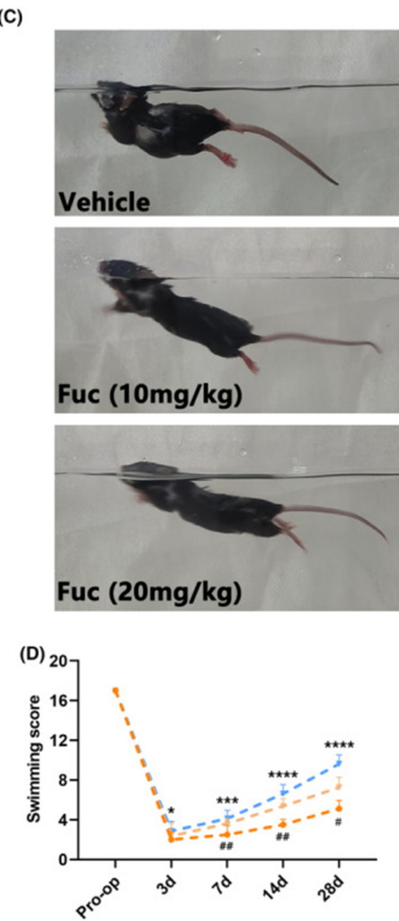
Swimming accurately reflects the motor ability of the nervous system of animals. Given the need for quantitative analysis, we performed multiple swimming tests at various time points after SCI, utilizing the LSS for assessment as illustrated in the quantitative data displayed in Figure 1 (panels C and D). As shown in Fig. 1D, the motor functions of all groups gradually recovered from the 7th day after SCI, and the recovery rate was significantly faster in the Fuc-treated groups, especially the 20 mg/kg group.
In the rotarod test performed on the 28th day after SCI, the Fuc-treated group showed better balance and motor coordination in all tests compared with the vehicle-treated group. In summary, the data obtained demonstrated a statistically significant improvement in motor function in the SCI mice as a direct result of Fuc administration, a conclusion supported by the synthesis of all experimental findings.
The inflammatory response plays a vital role in SCI treatment, and its regulation minimizes secondary damage and optimizes the local environment for repair. We used Iba1 as a marker to characterize microglia and macrophages, iNOS as a marker for M1-type inflammatory cells, and Arg1 as a marker for M2-type inflammatory cells. The study results clearly showed that Fuc intervention significantly limited the extent of inflammation within the injury site and significantly reduced the number of inflammatory cells. Fuc treatment reduced the expression of iNOS and simultaneously increased the expression of ARG1. The results showed that Fuc had a dual effect: not only did it suppress inflammation, but it also actively reversed the polarization state of the inflammatory cells involved. This suggests that Fuc not only limits the spread of inflammation and reduces the injury extent but also transitions inflammatory cells from a pro-inflammatory to an anti-inflammatory, pro-repair state.
The study used NeuN staining to count surviving neurons at varying distances from the injury site in coronal sections from each treatment group, to understand how functional recovery relates to the underlying anatomy. The number of surviving neurons in the Fuc treatment group was significantly higher than that in the vehicle group, and the neuroprotective effect of the Fuc 20 mg/kg group was better than that of the Fuc 10 mg/kg group. To further explore the benefits of Fuc in SCI repair, primary cortical neurons were treated with H2O2 (100 μM) for 24 hours to mimic neuronal injury. In an in vitro neuronal injury model, Fuc significantly enhanced neuronal survival, with a greater protective effect at higher concentrations.
The level of apoptosis was evaluated in mice seven days after SCI by performing TUNEL staining on their collected samples. The number of TUNEL-positive cells in the Fuc-treated group was significantly lower than that in the vehicle group. In contrast to the 10 mg/kg Fuc group, the 20 mg/kg Fuc group demonstrated a significantly enhanced anti-apoptotic effect. These results collectively confirm the neuroprotective role of Fuc in inhibiting neuronal apoptosis and promoting neuronal survival after SCI. This protective effect was more pronounced in the Fuc 20 mg/kg group than in the Fuc 10 mg/kg group.
Seven days following the induction of spinal cord injury in mice, TUNEL staining was utilized as a means of quantitatively evaluating the degree of apoptosis present in the collected tissue samples. The number of TUNEL-positive cells in the Fuc-treated group was significantly lower than that in the vehicle group. Furthermore, compared to the Fuc 10 mg/kg group, the Fuc 20 mg/kg group showed a stronger anti-apoptotic effect. These results collectively confirm the neuroprotective role of Fuc in inhibiting neuronal apoptosis and promoting neuronal survival after SCI.
In the study, they performed immunostaining of spinal cord sections using CC1 to identify mature oligodendrocytes and SOX10 to label cells within the OL lineage. Treatment with Fuc (20 mg/kg) also increased the proportion of CC1+ cells among SOX10+ cells. These findings show that Fuc boosts both the population of OL lineage cells and the maturation of OPCs.
A five-grade pathological scale for myelin sheath ultrastructure at the injury site was developed via TEM to accurately evaluate remyelination efficiency. Compared with the sham group, both the SCI + Veh and SCI + Fuc (20 mg/kg) groups showed various degrees of demyelination. However, Fuc treatment led to extensive remyelination (Fig. 2A). Notably, quantification of myelin pathology levels revealed that compared with the SCI + Veh group, the lesions in the SCI + Fuc group at 28 dpi had a higher percentage of normal myelin/abnormal lamellae and a lower percentage of inclusions/unfolded/myelin whorls. Taken together, these results demonstrate that Fuc treatment increases the number of axons in the injury area and strengthens the myelin sheath structure of axons.
The study revealed that Fucoidan enhances OPC maturation into oligodendrocytes by triggering the PI3K/AKT/mTOR signaling pathway. The data obtained from these experiments clearly demonstrate that fucoidan enhances spinal cord injury repair through a mechanism involving the modulation of the surrounding microenvironment and a consequential stimulation of remyelination processes.


Source: CNS Neurosci Ther. 2024 Aug; 30(8): e14903. .doi: 10.1111/CNS.14903
