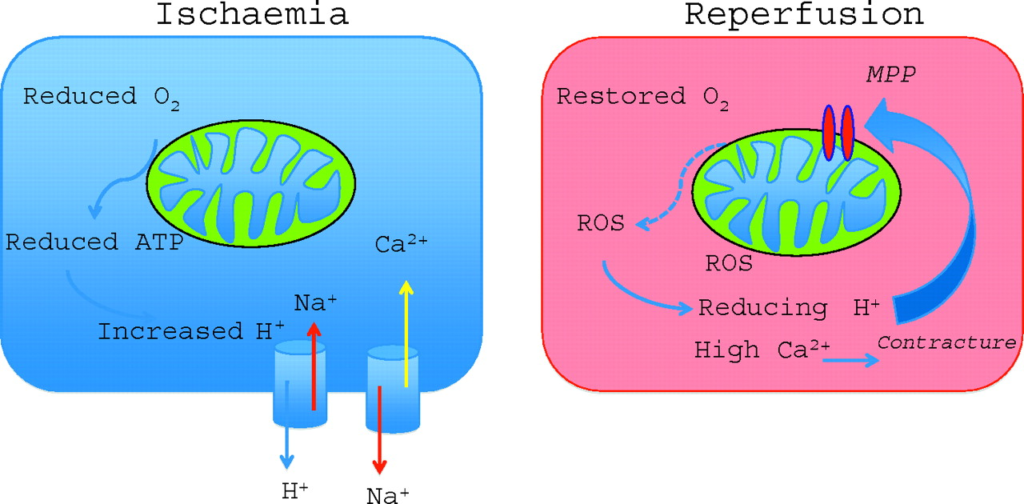
When myocardial infarction, also known as cerebral infarction, occurs, so-called ischemia-reperfusion injury occurs in which blood flow temporarily stops and resumes. In other words, when the blood flow stops, the oxygen supply to the tissue is cut off, and the tissue becomes hypoxic. As the blood flow resumes, oxygen is rapidly supplied, causing oxidative stress and destroying the cells.

This causes Ischemic stroke, a catastrophic neurological disorder, which is the third leading cause of death and permanent disability worldwide. Reperfusion into the ischemic brain is currently the best way to rescue the ischemic brain and limit infarction onset. Yet, the exact cause of brain IRI (Ischemia-Reperfusion Injury) is not fully understood.
Fucoidan has shown various beneficial activities, including anti-virus and anti-cancer effects. Hence, in this blog, I would like to share research on the effects of fucoidan on IRI in the brain and the underlying mechanism.
According to the study, “Protective Role of Fucoidan in Cerebral Ischemia-Reperfusion Injury through Inhibition of MAPK Signaling Pathway,” Nan Che et al. examined fucoidan’s effects on IRI in the brain.
First, using Sprague-Dawley (SD) rats, sham, IRI + saline (IRI + S), IRI + 80 mg / kg fucoidan (IRI + F80), and IRI + 160 mg / kg fucoidan (IRI + F160) ) was randomly administered to 4 groups. Fucoidan (80 mg/kg or 160 mg/kg) was intraperitoneally injected for seven days before the rats were inducted into the cerebral IRI model with the middle cerebral artery occlusion (MCAO) method.
As a result, it was observed that administration of 80 mg/kg fucoidan significantly reduced the infarct volume compared to the IRI + S group (p <0.01), and administration of 160 mg/kg fucoidan further significantly reduced the infarct volume. The IRI + S group (p <0.001) indicates that fucoidan may be dose-dependently protected from IRI (See Fig. 1).
And inflammation-related cytokines (interleukin (IL) -1β, IL-6, myeloperoxidase (MPO), tumor necrosis factor (TNF) -α), oxidative stress-related proteins (malondialdehyde (MDA), superoxide dismutase (SOD)) levels were measured in an ischemic brain by an enzyme-bound immunoabsorption assay (ELISA). The results indicated that IL-1β, IL-6, MPO, and TNF-α levels were significantly reduced by fucoidan treatment compared to IRI + S. It was also proven that fucoidan could substantially reduce the inflammatory response after ischemic injury of the brain in rats (See Fig.2).
In addition, levels of apoptosis-related proteins (p-53, Bax, Bcl-2) and mitogen-activated protein kinase (MAPK) pathways (phosphorylation-extracellular signal-regulated kinase (p-ERK), p-c-Jun N-terminal kinases. (JNK) and p-p38) were measured. The results showed that fucoidan administration significantly decreased all the p-p38, p-ERK, and p-JNK in a dose-independent way.
Thus, all results above further prove that fucoidan significantly reduced neurological defects and infarct volume in a dose-dependent manner in the IRI + S group. Fucoidan also significantly reduced the levels of inflammation-related cytokines, and oxidative stress-related proteins suppressed apoptosis and suppressed the MAPK pathway. Therefore, fucoidan plays a protective role in IRI in the brain and may be due to inhibition of the MAPK pathway.


