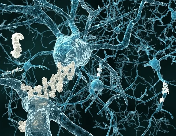
Alzheimer’s disease (AD) is the most common age-related neurodegenerative disease. It causes damage to the nervous tissue of the ability causes cognitive impairment such as depression and memory loss. Currently, there are some drugs available to suppress AD, but unfortunately, no effective treatment has been established yet. Amyloid plaque is one of the most common pathological features in the brains of AD patients. Amyloid plaque is composed of amyloid β (Aβ).
If an abnormal protein larger than average amyloid β is formed, it accumulates instead of getting eliminated. A few studies mention that it begins to accumulate in the brain at least 20 years before the onset of dementia. The accumulated amyloid β is thought to kill brain cells. It is also considered that forgetfulness occurs when the brain cells, the subject of memory, die. In a few cases, Amyloid β can also deposit in blood vessels and can cause cerebral hemorrhage.

However, the accumulation mechanism has not been completely explained up till now. Despite that, it is said that highly toxic amyloid β begins to accumulate when decomposition and elimination do not go well due to aging.
On the other hand, there is some hope through Fucoidan, a sulfated polysaccharide in brown algae. In recent studies, it has been reported to have a neuroprotective effect. It is also expected to have therapeutic and preventive effects on neurodegenerative diseases such as Alzheimer’s.
Therefore, in this blog, I would like to introduce using AD model nematodes to examine the impact of Fucoidan on Aβ-induced neurotoxicity from the viewpoint of proteasome activity and active oxygen production. This study is conducted by Xuelian Wang et al., “Fucoidan inhibits amyloid-β-induced toxicity in transgenic Caenorhabditis elegans by reducing the accumulation of amyloid-β and decreasing the production of reactive oxygen species. “
First, Caenorhabditis elegans, a nematode genetically modified to produce Aβ when the growth temperature rises from 16³C to 25³C, was used as an AD model nematode. The state of Aβ deposition was compared between the fucoidan-administered and the non-administered groups. Aβ of nematodes was stained with thioflavin S, and the deposited Aβ was observed. As a result, the fucoidan-administered group (500 ng / mL) decreased by about 47% after 60 hours and about 52 after 1018 hours compared with the non-administered group. It was proven that Fucoidan significantly inhibited the deposition of Aβ (Fig. 1).
Next, we examined the proteasome activity, a protein complex involved in the removal of Aβ, in a similar AD model nematode. As a result, the proteasome activity in the fucoidan-administered group increased by 2 and 3 times, respectively, in both concentrations of 100 and 500 ng / mL compared with the non-administered group (Fig. 2).
Thus, this experiment suggested that proteasome activity is associated with the suppression of Aβ deposition in Fucoidan. Finally, we examined oxidative stress caused by active oxygen, which is proven to be one of the significant causes of neurotoxicity caused by Aβ. As a result of measuring the amount of active oxygen produced in nerve cells in AD model nematodes grown under the same conditions, it is confirmed that Fucoidan suppressed the increase in active oxygen production due to Aβ induction by about 80% (Fig. 3). Hence, this study suggests that Fucoidan suppresses Aβ deposition by activating the proteasome and concomitantly suppresses the production of reactive oxygen species, by this means inhibiting neurotoxicity. In the future, research of Fucoidan on dementia at the clinical trial level is expected.


