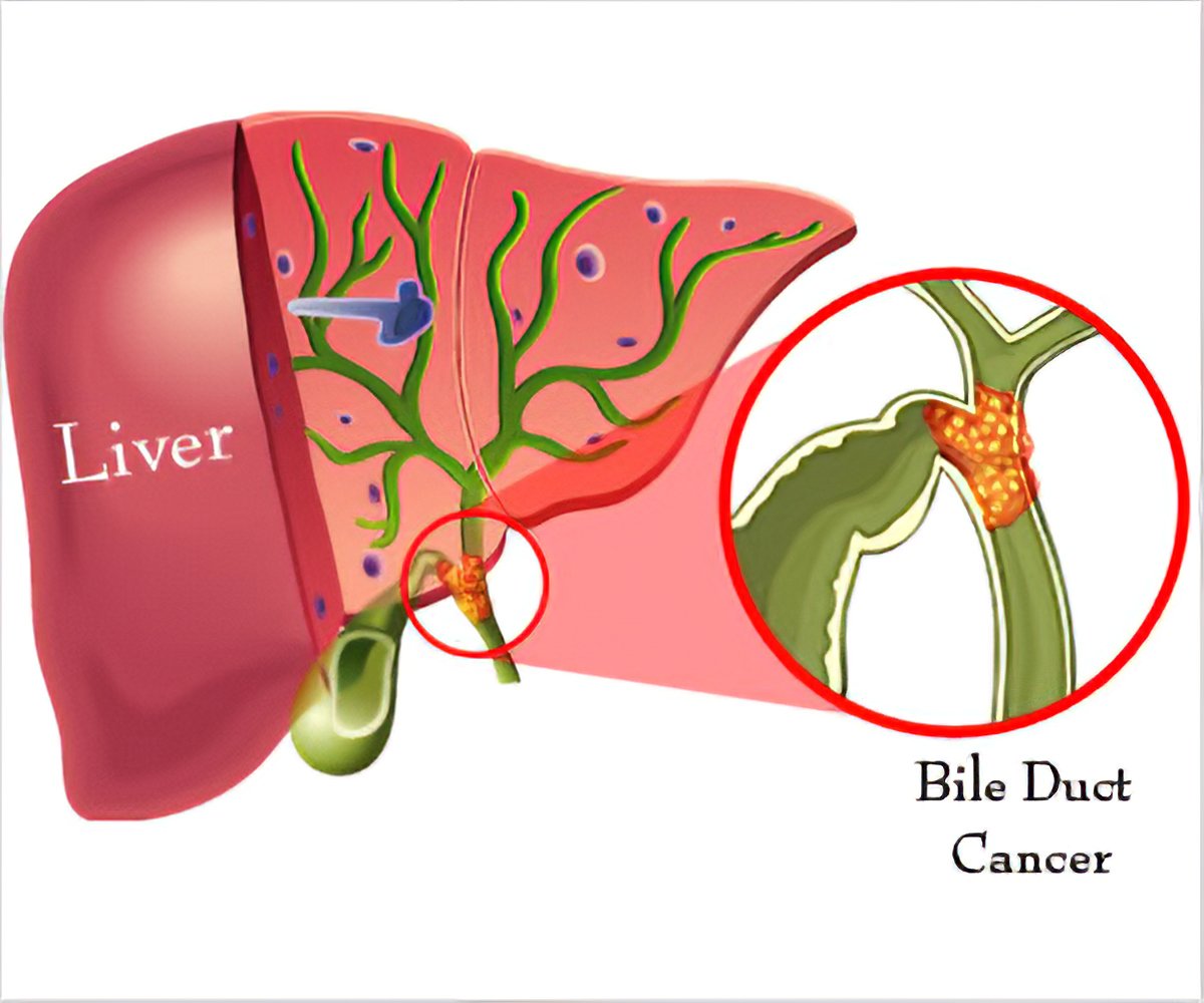
Bile duct cancer is a highly malignant form of cancer. It begins in the bile duct epithelial cells inside and outside the liver and is also known as Cholangiocarcinoma. Although the incidence of bile duct cancer is low in many countries, it has been reported that the incidence is high in Southeast Asian countries, especially Laos, Cambodia, and northeastern Vietnam.
In addition, as the symptoms of bile duct cancer appear late, there is no means for early diagnosis. Hence, sadly most patients with bile duct cancer that has metastasized have a 5-year survival rate of 10% or less.

On the other hand, Fucoidan, a natural high molecular weight polysaccharide with a sulfate group obtained from brown algae, contains various beneficial physiological activities. Significantly, the anticancer effect is attracting attention.
So, in this blog, I would like to share a fucoidan and bile duct cancer study based on the research content of “Anticancer Activity of Fucoidan via Apoptosis and Cell Cycle Arrest ” Cholangiocarcinoma Cell” by Pathanin Chantree et al.
Thus, researchers examined the anticancer effect of Fucoidan derived from Fucus vesiculosus on the bile duct cancer cell CL-6. First, they had examined the cell viability of CL-6 cells supplemented with Fucoidan and OUMS, a normal human cell. Then, Fucoidan was added to each at concentrations of 0, 100, 200, 300 µg / mL and measured 24, 48, 72 hours later. As a result, it had almost no effect on the viability of OUMS cells. In contrast, CL-6 cells of the cell viability decreased in a concentration-dependent manner (Fig. 1).
Additionally, to investigate the apoptosis-inducing effect on CL-6 cells, the cells were cultured for 48 hours in the presence of Fucoidan at the same concentration and then analyzed with a flow cytometer. As a result, many cells that died due to apoptosis were detected in CL-6 cells treated with Fucoidan, but few dead cells were detected in OUMS cells.
Next, they had investigated the effect of Fucoidan on the cell cycle of CL-6. Forty-eight hours after fucoidan treatment, analysis was performed with a flow cytometer using PI (propidium iodide), which detects DNA replication. As a result, it was indicated that the number of cells in the G0 / G1 phase increased depending on Fucoidan concentration, that Fucoidan increased the cell cycle and stopped the cell cycle.
Furthermore, when the expression of apoptosis-related proteins was examined by Western blotting, the expressions of cleaved PARP, apoptosis-promoting Bax, and apoptosis-inducing cleaved caspase-3 were found at the beginning of apoptosis increased in a concentration-dependent manner. Besides, the expression of Bcl-2, which suppresses apoptosis, decreased in a concentration-dependent manner (Fig. 2). The above results clarified that Fucoidan effectively executes an anticancer effect on bile duct cancer cells through cell cycle arrest and apoptosis-inducing action.
On a positive note, it is expected to expand into research on fucoidan biological effects such as clinical trials in the future.


