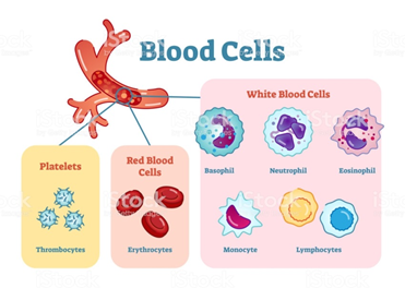
Blood cells also include white blood cells. These white blood cells play a significant role in eliminating foreign substances such as bacteria and viruses that have entered the body from the outside. Red blood cells carry oxygen, and platelets are tiny blood cells that stop bleeding by forming clots. The origin of these blood cells is called hematopoietic stem cells, which are present in the bone marrow inside the bone. They differentiate while proliferating (immature cells become mature cells) and become blood cells. Hematopoietic stem cells are divided into myeloid stem cells and lymphoid stem cells. The myeloid stem cells produce erythrocytes, platelets, neutrophils, and monocytes. The lymphoid stem cells produce lymphocytes such as B cells, T cells, and NK cells. Neutrophils, monocytes, and lymphocytes are classified into white blood cells and make up their significant components.
In this blog, I will discuss in detail of chronic myelogenous leukemia and fucoidan effect.
According to the Leukemia and Lymphoma Society, by the end of 2020, the estimated cases of leukemia in the USA will reach 60,530. Moreover, in 2019, 22,840 people were expected to die from leukemia (13,150 males and 9,690 females).
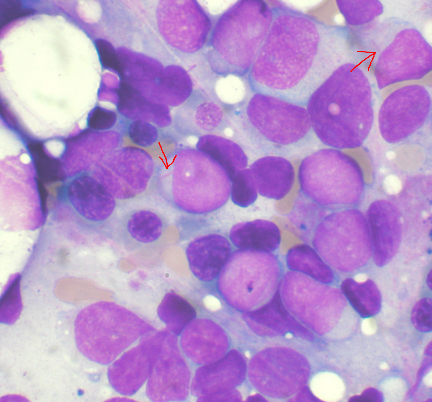
Chronic myelogenous leukemia develops when there are abnormalities in hematopoietic stem cells, and cancerous blood cells proliferate indefinitely. The most noticeable defect in the test value is an increase in white blood cells. However, at the same time, anemia and an increase in platelet count may also be observed. Chronic myelogenous leukemia is one of the relatively slow progression types of blood cancer.
As for the symptoms, chronic myelogenous leukemia has almost no symptoms at an early stage. The primary reason is that when white blood cells become cancerous and turn into leukemia cells, they function almost like healthy white blood cells and progress slowly. Therefore, it is pointed out that the number of white blood cells is increased in medical examinations, and more than half of them are found by accident.
On the other hand, as the disease progresses, white blood cell count, platelet count, and anemia gradually appear. As the white blood cell count increases, the following symptoms are observed: general fatigue and lethargy, night sweats, weight loss, and bloating of the abdomen due to an increase in the spleen size.
Most of the causes of onset are that the Philadelphia chromosome (Ph) is generated by exchanging a part of chromosome 9 and 22. An abnormal gene called the BCR-ABL fusion gene is formed on the Philadelphia chromosome. A protein made from the BCR-ABL fusion gene creates excess blood cells, which leads to chronic myelogenous leukemia.
According to the research of “Anticancer activity of Sargassum Oligocystum (brown algae) water extract against human cancer cell lines” by K. Zandi et al., the most effective antitumor activity has been shown at concentrations 500 µg/ml and 400 µg/ml of the alga extract against Daudi (Burkitt’s lymphoma) and K562 (Chronic myelogenous leukemia) cell lines, respectively. The results showed that the extracts of brown alga Sargassum Oligocystum have remarkable antitumor activity against K562 and Daudi cell lines. I believe this is justified and can indeed be suggested for further research such as algal extract fractionation and purification and in vivo studies to formulate natural compounds with antitumor activities. Also, according to Ichiro Teruya et al., the analysis of “Anticancer effects of enzyme-digested fucoidan extract from seaweed Mozuku.”, chronic myelogenous leukemia K562 cells was suppressed the proliferation by the fucoidan extract treatment.
Results of Viability Test for K562 cell line after treatment with algal extract
Number of dead cells Number of viable cells Extract concentration (µg/mL)
1.3 × 10⁵ 10.2 × 10⁵ 0
1.2 × 10⁵ 9.1 × 10⁵ 100
1.2 × 10⁵ 7 × 10⁵ 200
1.5 × 10⁵ 6.9 × 10⁵ 300
1.5 × 10⁵ 4.5 × 10⁵ 400
1.6 × 10⁵ 4.6 × 10⁵ 500
The most effective concentration in which the number of viable cells was decreased significantly was 500 µg/ml of algal extract. Dead cells were presented in all concentrations of the algal extract. The most amount of dead cell has been shown for 400 and 500 µg/ml of algal extract.