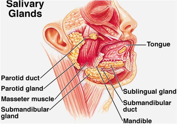
The function of saliva is to help digest food, and chewing well helps prevent overeating. It also has a cleansing action of washing away food debris and keeping the mouth clean, neutralizing the oral cavity, and preventing tooth decay. Researchers have discovered many bacteria are in the mouth, and substances such as lysozyme and lactoferrin in saliva prevent bacteria growth.
In addition, peroxidase contained in the saliva is said to have the function of suppressing the toxicity of carcinogenic substances. Thus, saliva has an important role, but in treating thyroid cancer, radioactive iodine (RI) administration is essential to remove the cancer cells and prevent cancer from recurring. RI not only affects thyroid tissue but also penetrates other tissues such as salivary glands (SG), breast, and gastrointestinal tract, causing unwanted side effects. Also, treating Radioactive iodine (RI) can destroy cellular components of salivary glands (SG). Although amifostine is the only radio-protective drug that scavenges ROS, it has various side effects and is quite expensive. On the other hand, Fucoidan is extracted from seaweed and shows a wide range of biological activities such as anti-inflammatory, anti-virus, and anticancer effects.

So, in this blog, I would like to introduce the “Fucoidan attenuates radioiodine-induced salivary gland dysfunction in mice” study by Young-Mo Kim et al. This study investigated whether Fucoidan could alleviate the SG dysfunction induced by radioactive iodine.
First, thirty-six mice were divided into three groups (n = 12/group): standard control group, RI-treated group (0.01 mCi/g body weight), Fucoidan (6 hours before intraperitoneal RI exposure and two doses of 10 mg/kg 0.5 hours before). In each group, all mice were given 1.5 μg thyroxine (T4)/100 g body weight and 1% calcium lactate in their drinking water to maintain euthyroid status.
There was no significant difference in body weight between groups before the start of the experiment. However, after the investigation, the mice in the RI group weighed significantly less than the normal control group mice at the mean±s.d. Mean ± SD at 2, 4. At 12 weeks, mice in the fucoidan group were significantly heavier than mice in the RI group at 2 and 12 weeks after treatment (See Fig. 1 b). For salivary lag time and salivary flow rate, statistical analysis used the Kruskal-Wallis test and Dunn’s post hoc multiple comparison test, and gland weights were not remarkably different between all groups (See Fig. 1c). The lag time and salivary flow in the fucoidan group were shorter and more significant than in the RI group (See Fig. 1 d)
Early-stage injury induced by RI is characterized by painful swelling of the SG. This swelling is associated with poor oral intake and weight loss. In the later stages of RI therapy, SG function continues to deteriorate, and patients complain of dry mouth and significant discomfort. The fucoidan-treated group improved these functional parameters (See Fig.1 a).
Acinar atrophy, inflammation, low mucin staining parenchyma, and ductal fibrosis are the main histopathological features of RI-induced SG injury, although fibrosis induces ductal constriction. It is a factor that causes secretion disorders. The results showed that histological features were improved in the fucoidan-treated group compared with the RI group (See Fig.2)
RI destroys SGs primarily by damaging cellular components and initiating apoptosis. Immunohistochemical staining showed that the expression of AQP5 and α-SMA was higher in the fucoidan group compared with the RI group. These findings suggest that Fucoidan can protect salivary epithelial and myoepithelial cells from RI-induced cell damage.
The study shows that fucoidan administration attenuates RI-induced SG damage in mice before RI exposure. We believe that Fucoidan should be considered a candidate for preventing SG dysfunction in thyroid cancer patients undergoing RI therapy.


