
Previous studies have confirmed the impact of fucoidan extracted from Sargassum hemiphyllum on various HCC cell types (such as Huh6, Huh7, SK-Hep1, and HepG2) in both in vivo and in vitro settings, but the exact molecular events underlying these effects have yet to be fully understood. So in this blog, I would like to share the following first study, “Fucoidan Elevates MicroRNA-29b to Regulate DNMT3B-MTSS1 Axis and Inhibit EMT in Human Hepatocellular Carcinoma Cells (HCC)” by Ming-De Yan et al. According to the research, fucoidan plays a role in regulating miR expression, leading to the activation of the tumor suppressor cascade in HCC cells.
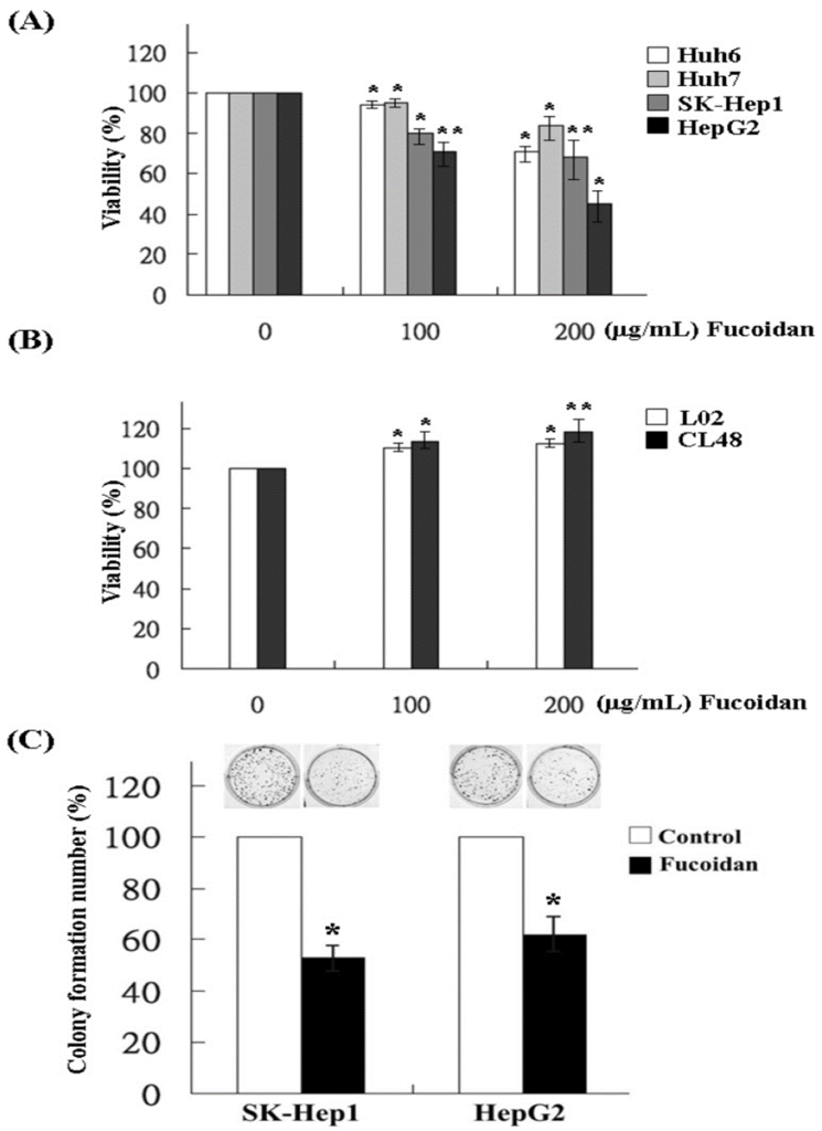
In order to assess the influence of fucoidan on HCC cell proliferation, we conducted MTS assays on Huh6, Huh7, SK-Hep1, and HepG2 cells. After 48 h of fucoidan treatment, various degrees of proliferation inhibition were observed in HCC cells. The inhibition of cell proliferation in Huh6, Huh7, SK-Hep1, and HepG2 cells was observed when exposed to fucoidan at a concentration of 200 μg/mL, as shown in Figure 1A. In contrast, the results indicated that the proliferation of normal human liver cell lines remained unaffected when treated with fucoidan at the same dosage. This observation strongly implies that fucoidan exhibits a preferential inhibitory effect on cancer cells, as highlighted in Figure 1B. We next examined the effect of fucoidan on clonogenicity in SK-Hep1 and HepG2 cells, which are relatively more sensitive to fucoidan than the other two HCC cell lines. The colony formation of SK-Hep1 and HepG2 cells was significantly reduced when treated with fucoidan at a dose of 200 μg/mL, as indicated by the results in Figure 1 C.
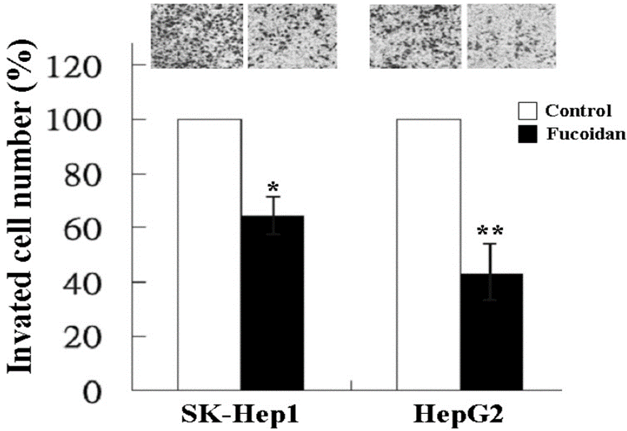
The transwell invasion assay was used to investigate how fucoidan affects the invasion of SK-Hep1 and HepG2 cells. As shown in Figure 2, at a dose of 200 μg/mL, fucoidan significantly reduced the invasion of SK-Hep1 and HepG2 cells.
As miR regulation has become a new approach for cancer therapy, the miR expression profile of fucoidan-treated HepG2 cells was analyzed by Affymetrix GeneChip miRNA 2.0 Array. The results showed that fucoidan increased the expression of tumor-suppressing miRs, such as miR-29 family and miR-1224.
The increase in miR-29b expression by fucoidan was further confirmed by real-time quantitative PCR. miR-29b expression in fucoidan-treated normal cell line (L02) and HCC cells was all significantly increased in a dose-dependent manner. In comparison to L02 cells, SK-Hep1 and HepG2 cells displayed significantly decreased basal levels of miR-29b. Furthermore, the induction of miR-29b by fucoidan appeared to be more pronounced in these two HCC cell lines.
As a result of predicting the targets of miR-29b, DNMT3B showed the highest score and was found to be a potential miR-29b target gene. Then, they verified the suppression of DNMT3B by miR-29b in HCC cells. Following the significant upregulation of miR-29b expression caused by fucoidan, the luciferase activity of the wild-type DNMT3B 3′-UTR reporter was noticeably reduced in SK-Hep1 and HepG2 cells treated with fucoidan for 48 hours. However, there was no suppression observed in the luciferase activity of the mutant DNMT3B 3′-UTR reporter. Similar compatible results were observed in these two types of HCC cells transfected with miR-29b mimics, indicating that fucoidan increased miR-29b expression and negatively regulated DNMT3B in HCC cells.
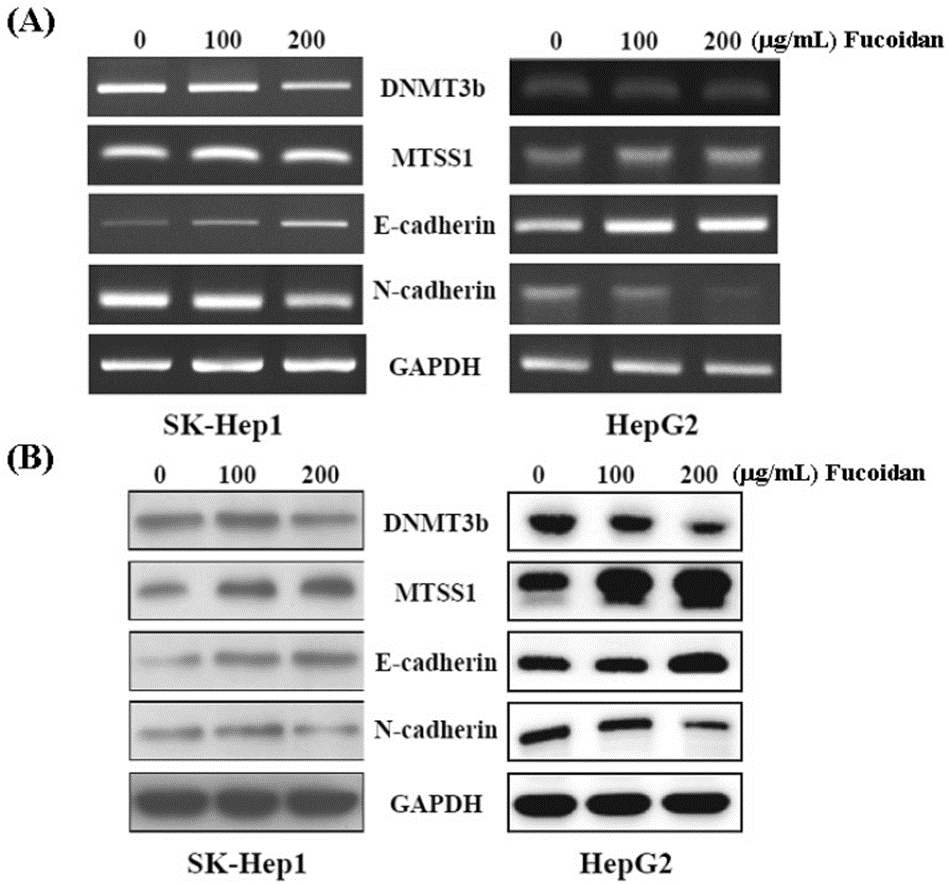
To confirm the inhibitory effect of fucoidan on DNMT3B, we performed western blot and RT-PCR. As shown in Figure 3, both the mRNA and protein levels of DNMT3B were dose-dependently suppressed in fucoidan-treated SK-Hep1 and HepG2 cells. The levels of MTSS1 mRNA and protein were consistently elevated in a dose-dependent manner by fucoidan, while the expression of DNMT3B was suppressed in these HCC cells. Also, the increase in E-cadherin and the inverse decrease in N-cadherin represent the inhibition of EMT in these fucoidan-treated HCC cells.
Fucoidan also suppressed TGF-β signaling in these HCC cells. The administration of fucoidan for 48 hours resulted in a dose-dependent reduction of TGF-β receptor 1 and 2 (TGF-βR1, 2), phosphorylated Smad2/3 (p-Smad2/3), and Smad4 protein levels, accompanied by an increase in the inhibitory Smad protein, Smad7. They then further investigated its effects on MMPs and TIMPs. Fucoidan dose-dependently decreased the protein levels of MMP2 and MMP9 and increased the level of tissue inhibitor of metalloproteinase 1 (TIMP-1). These effects may also contribute to the reduced invasive activity of HCC cells shown in Figure 2.
Fucoidan blocks TGF-β signaling and effectively controls the miR-29b-DNMT3B-MTSS1 pathway, resulting in the suppression of EMT, prevention of extracellular matrix breakdown, and inhibition of migration and invasion in HCC cells.
The results in this study show that fucoidan increased miR-29b and suppressed DNMT3B, resulting in the upregulation of MTSS1. Meanwhile, fucoidan downregulated the TGF-β signaling pathway in HCC cells. These effects led to the inhibition of EMT and the prevention of extracellular matrix degradation, reducing the invasive activity of HCC cells. These results indicate that fucoidan has promising potential as a holistic treatment option to enhance the treatment results for patients with HCC.
Source: Mar Drugs. 2015 Oct; 13(10): 6099–6116. doi: 10.3390/md13106099