
With recent advanced studies, Fucoidan has shown various beneficial biological functions. Fucoidan inhibits cancer cell proliferation and inhibits cell proliferation by inducing apoptosis and cell cycle arrest. Previous studies have also indicated that Fucoidan is a potential preventive or therapeutic agent for cancer, suggesting that tumor progression is influenced by the microenvironment, including inflammatory responses.
Cell density has been demonstrated to be related to environmental factors such as inhibitors, substrate accumulation, and depletion of essential nutrients or serum growth factors. However, the specific mechanisms underlying Fucoidan’s anticancer effects on the cellular environment are yet to be clarified.
Hence, in this blog, I would like to share the study “Differences in cell death and cell cycle following fucoidan treatment in high-density HT-29 colon cancer cells” by IN-HYE KIM et al.
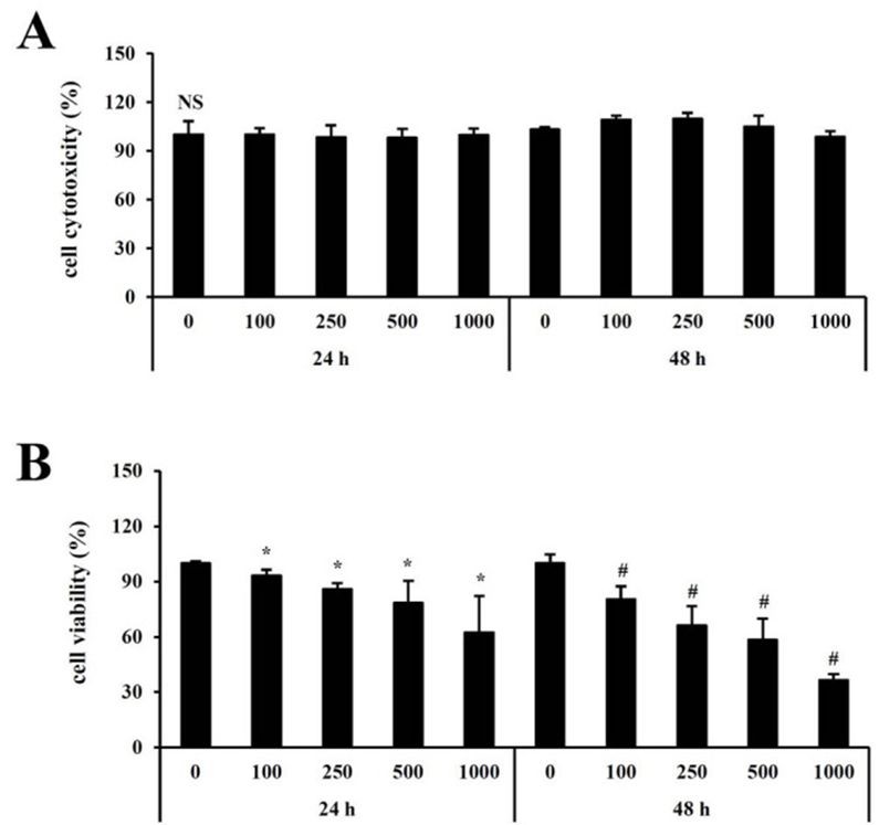
Previous studies used normal-density cells but examined the effect of fucoidan treatment on colon cancer cell lines HT-29 cultured at high density. Cell viability and cytotoxicity were investigated, and no cytotoxicity was detected with fucoidan treatment at any concentration (0-1,000 μg/ml) or time points (24 and 48 hours). (See Figure 1 A) Cell viability was significantly suppressed after fucoidan treatment compared to untreated cells. (See Figure. 1B). Notably, the viability decreased to 50% at a 500 µg/ml concentration when treated with Fucoidan for 48 hours.
Nuclear morphological changes were also examined to provide direct evidence of apoptosis by fucoidan treatment. Apoptotic bodies were detected by nuclear-specific DNA fragmentation. Therefore, inhibition of HT-29 cell viability by fucoidan treatment was closely associated with the induction of apoptosis and DNA synthesis.
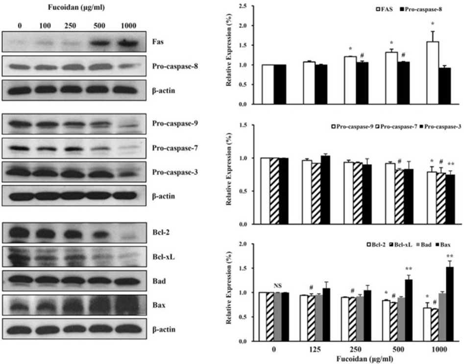
Furthermore, they investigated the effect of fucoidan treatment on the apoptotic properties of high-density HT‑29 cells. Receptors containing FAS are essential in the physiological adjustment of apoptosis. These factors and functions cause apoptosis as a defense against diseases caused by various tumors and immune systems. The investigation included the external genital route through the high-density HT-29 cells processed in the Fucoidan and the protein expression level of the FAS and Caspase-8.
The result shows that the fucoidan treatment increased the expression of the FAS protein, reducing the expression of Caspase -8 proteins. (See Figure. 2) Further survey of endogenous routes using Caspase-9, -7, and -3 protein expression levels of high-density HT-29 cells treated with Fucoidan. When processed with the reagent, Caspase-9, -7, and -3 protein expression levels decreased. (See Figure. 2).
They studied whether Fucoidan affects cell survival (BCL-2 and BCL-XL) and cell death (BAD and BAX) in the Signal Transmission of the BCL-2 family. As a result, the fucoidan treatment increased the expression of the BAX protein as concentration-dependent, reducing the expression amount of BCL-2 and BCL-XL. (See Figure. 2)
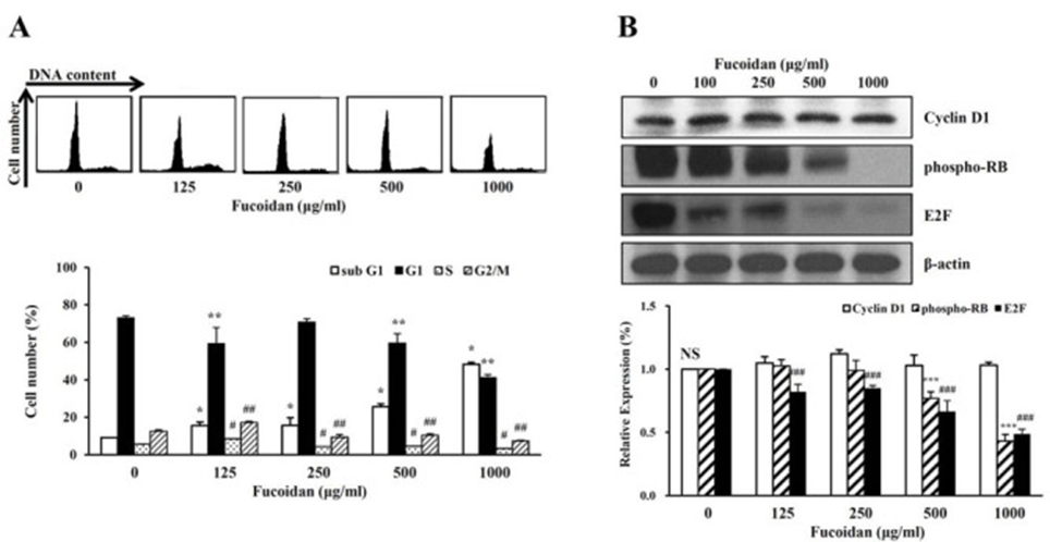
It also indicated that high-concentration Fucoidan triggers a morphological change, including a decrease in cell survival rate and a fragment of nuclear weapons. Thus, the characteristics of apoptosis in the cell cycle of high-density cells processed in Fucoidan were examined. The number of cells in the sub-G1 phase increased compared to unprocessed cells when treated with Fucoidan. (See Figure. 3A) Contrarily, the number of cells in the G1, S, and M phases decreased to unprocessed cells.
Next, the expression level of protein that regulates the cell cycle of the G1 period was investigated. Cycline D1 protein expression levels were not affected by fucoidan treatment. The phosphorylated level of RB decreased dose-dependent with fucoidan treatment. The E2F protein was inhibited by binding the fluorescent RB in the G1 term. E2F protein expression levels have dropped by fucoidan processing. (See Figure. 3B) These results suggest that the reduced DNA synthesis hindered the phosphorylation of RB proteins by creating complexes between E2F and RB proteins. Hence, when processed with a high -concentration of Fucoidan, the G1 phase stopped, and the cell growth (cell survival rate) was suppressed.
The relationship between the cell environment and the functional effect of Fucoidan is expected to develop Fucoidan as a potential treatment for future research and cancer treatment.
Source: Mol Med Rep. 2017 Jun; 15(6): 4116–4122 doi: 10.3892/mmr.2017.6520