NAFLD, which stands for nonalcoholic fatty liver disease, is a widespread condition that affects approximately 25% of the global population. It is important to note that the progression of NAFLD is strongly linked to liver failure, insulin resistance, and cardiovascular disease.
So, in this blog, I would like to share the study “Antioxidant and Antisteatotic Activities of Fucoidan Fractions from Marine and Terrestrial Sources” by Zeinab El Rashed et al. The fucoidan fraction extracted from the brown alga Cystoseira compressa (CYS) and the fucoidan fraction found in the terrestrial tree Eucalyptus globulus (EUC) have been found to possess antioxidant and antisteatogenic effects. In this discussion, we will delve into the chemical and structural features of both CYS and EUC, as well as their utilization in vitro assays and experimental cell models. These studies are particularly relevant to exploring their potential benefits in addressing oxidative stress-related metabolic diseases, including NAFLD and atherosclerosis.
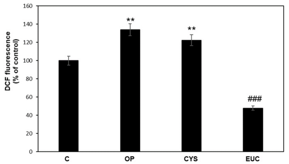
First, antioxidants can be useful as therapeutic compounds against oxidative stress-related diseases. Hence, they chose to assess the impact of CYS and EUC (fucoidan extracted from Eucalyptus, land plants) on antioxidants in cellular systems. The antioxidant capacity of CYS and EUC was analyzed using a 2′,7′-dichlorofluorescein diacetate (DCF-DA) assay for ROS detection in FaO (hepatoma) cells (see Figure 1). OP treatment significantly increased ROS production, but it was not significantly affected by CYS treatment. On the contrary, EUC was able to reduce ROS production in OP cells even below control values. Oxidative stress is believed to initiate liver damage and is believed to contribute to the advancement of NAFLD and the development of more severe pathological conditions.
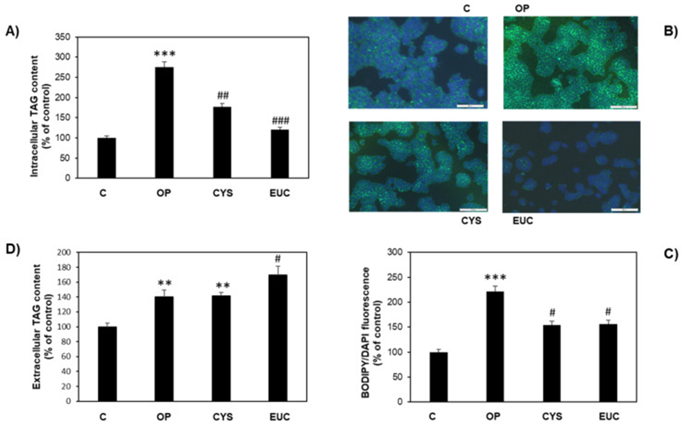
Next, they investigated whether antioxidant effects were associated with antisteatogenic effects in lipid-overloaded FaO cells. Intracellular triacylglycerol (TAG) content was quantified in both control cells (C) and adipocytes in the absence (OP) or presence of CYS or EUC using two different methods (see Figure 2A). Incubation with 50 μg/mL CYS or EUC during OP treatment decreased TAG content by 35% and 56%, respectively, compared to OP. The hypolipidemic properties of the two FU extracts were confirmed by BODIPY/DAPI staining of FaO cells. They were able to visualize intracellular lipid droplets (LDs) stained green with BODIPY 493/503 (Figure 2B). The fluorescence levels were measured using spectrofluorimetry and the resulting data was used to generate the bar plot depicted in Figure 2C.
Additionally, TAG secretion by FaO cells was studied by measuring His TAG content in the medium of the same cells. The results depicted in Figure 2D clearly demonstrated a significant increase in TAG secretion in OP cells, as compared to the control group. His subsequent CYS treatment did not affect this value, whereas EUC treatment significantly enhanced this secretion to OP.
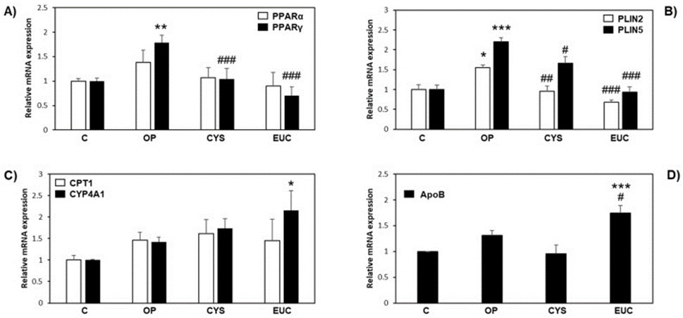
Peroxisome proliferator-activated receptors (PPARs) are ligand-activated transcription factors that play an important role in NAFLD. PPARα and PPARγ represent the most abundant PPAR isoforms expressed in FaO cells. Hepatic steatosis is associated with the expression of PPARγ, a well-known marker that plays a role in activating genes related to adipogenesis and regulating perilipin (PLIN). This induction is likely to occur in FaO cells to enable efficient energy storage. Figure 3A shows that only PPARγ expression was significantly induced by HisOP treatment and was downregulated to control levels after incubation with both CYS and EUC. The regulation of PLIN2 and PLIN5 expression, which is controlled by PPARγ, resulted in corresponding changes.
Indeed, steatotic FaO cells showed a significant increase in both PLIN2 and PLIN5 mRNA levels compared to controls. In contrast, the treatment with CYS or EUC resulted in a considerable decrease in expression, as shown in Figure 3B, in comparison to OP. As shown in Figure 4C, genes encoding proteins involved in lipid catabolism, such as carnitine palmitoyltransferase I (CPT-1) and cytochrome P450 CYP4A1, did not change their expression levels under different experimental conditions, except for cytochrome CYP4A1. It was. EUC significantly increased the levels of P450 enzymes, which are responsible for microsomal fatty acid ω-oxidation, compared to the control group.
Figure 4D shows that EUC was the only treatment that could induce the expression of ApoB mRNA. ApoB is the major apolipoprotein of liver VLDL. Hence, this finding suggests that EUC can enhance the release of liver fat, aligning with the observed rise in extracellular TAG in the same experimental settings (see Figure 3D). The findings show that CYS and EUC are responsible for reducing the intrahepatic energy storage mechanisms and altering the transport of LD. The mobilization of excess fat from LDs in EUC appears to be managed by microsomal ω-oxidation and the secretion of TAG through VLDL synthesis, resulting in a powerful anti-fat accumulation effect on liver tissue.
According to the DCF assay, both CYS and EUC effectively decreased ROS production to levels comparable to the control. The application of OP treatment led to a notable boost in NO production in HECV cells when compared to the control group. However, this increase was effectively mitigated by EUC treatment. It was observed that the increase in intracellular TAG content caused by OP treatment was significantly reduced only when EUC incubation was implemented.
As a result, two fucoidan fractions from both marine (CYS) and terrestrial (EUC) sources exhibited radical scavenging activity, antioxidant properties, and antilipolytic activity. The findings of the research showed that terrestrial fucoidans, particularly the EUC, exhibited stronger effects when compared to CYS. This suggests that these compounds could be valuable in the fight against NAFLD and its associated diseases.
Source: Molecules. 2021 Aug; 26(15): 4467. doi: 10.3390/molecules26154467