In my previous blog, I discussed the fucoidan-anticancer effect on hepatocellular carcinoma. Now, in this article, I want to share additional details based on the research of “Anticancer effect of fucoidan on cell proliferation, cell cycle progression, genetic damage and apoptotic cell death in HepG2 cancer cells” by P. Arumugam et al.
This extensive research will determine, anticancer effect by evaluating cell viability, colony formation, cell migration, cell cycle progression, genetic damage, and apoptosis. Thus, helping to consider the actual impact based on the results.
Firstly, an in-depth study was conducted on the influence of Fucoidan on HepG2 and the cells’ survival rate. Results of adding Fucoidan in fucoidan concentration form to HepG2. During the examination, there was a significant decrease in the survival rate of HepG2 in a concentration-dependent manner. Thus, proving the survival rate with the help of fucoidan concentration.
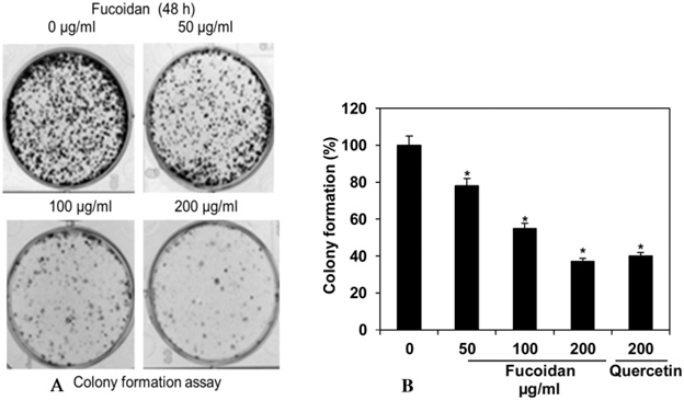
Next, Fucoidan on HepG2 cancer cells’ clonogenic effect was analyzed by colony-forming assay. HepG2 was sown on a plate, and the colonies (cell aggregates) were observed to investigate the tumorigenic state. As a result, the number of HepG4 colonies decreased as the fucoidan concentration increased.
These results indicate that Fucoidan has a tumorigenicity-suppressing effect on HepG2. (Fig.1)
Additionally, cell migration is one of the essential processes between cancer cells’ development and the spread of metastases. A wound-healing assay investigated Fucoidan’s inhibitory effect on HepG2 cancer cells’ migration. They examined how scratching the single layer of HepG2 and how the proliferation and migration of HepG2 filled the wound. As a result, it was found that the addition concentration of Fucoidan gradually suppressed cancer cell migration and growth.
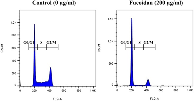
Moreover, investigating the cell cycle, when the HepG2 cell division process added Fucoidan, cell division in the G0/G1 phase (condition of cell division stopped) was increased. And the S phase (DNA’s replication condition) and the G2 / M phase in which the cells divide (Fig. 2). The number of cells was decreasing significantly. It was also found that Fucoidan arrests HepG2 cell division and suppresses HepG2 proliferation.
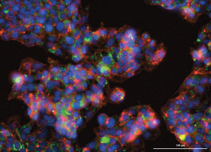
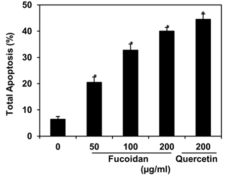
Regarding HepG2 apoptosis, Fucoidan was shown to damage the DNA in the cell nucleus of HepG2 and increased apoptotic cells (Fig. 3). In conclusion, it was proved that Fucoidan showed a promising anticancer effect on HepG2 by inducing DNA damage and apoptosis of HepG2 and suppressing the proliferation and migration of HepG2.
Reference: https://www.ncbi.nlm.nih.gov/pmc/articles/PMC6587026/