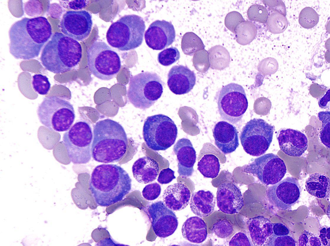
Multiple myeloma (MM) is a lymphocyte among white blood cells that differentiate from B cells (immature cells become mature cells). It becomes cancerous, thus evolving into myeloma cells. It is a disease in which myeloma cells increase mainly in the bone marrow. Still, cancerous myeloma cells continue to produce antibodies (M protein) that are incapable of attacking foreign substances.

Also, due to increased myeloma cells and M protein, various symptoms such as high calcium concentration in blood (hypercalcemia), renal failure, anemia, low back pain due to spine fracture, and uncomplicated fracture (bone lesion) occur in the body. In MM, angiogenesis around the tumor is believed to affect the progression and prognosis of the disease.
While there are treatments such as thalidomide and lenalidomide, new therapies are much needed because they are challenging to treat due to drug resistance. Studies on the angiogenesis-suppressing effect of fucoidan, a slimy component of brown algae with antitumor effects, using human umbilical vein endothelial cells have been reported. However, the impact on MM has not been clarified yet.
Therefore, in this blog, I would like to share the research “Fucoidan inhibits angiogenesis induced by multiple myeloma cells” using cultured cells and mice on the inhibitory effect of fucoidan on the induction of angiogenesis caused by MM.
First, it was reported that multiple factors work to induce angiogenesis in cancer cells. In the case of MM, hypoxia-inducible factor (HIF-1α) and vascular endothelial growth factor (VEGF) are the main factors. It is known to be an inducing factor. Expression of HIF-1α and VEGF is regulated by phosphatidylinositol 3-kinase (PI3K / AKT) and extracellular signal-regulated kinase (ERK1 / 2, p-ERK1 / 2).
In the case of MM, hypoxia-inducible factor (HIF-1α) and vascular endothelial growth factor (VEGF) are the main factors known to be inducing factors. As a result, the expression levels of HIF-1α and VEGF decreased in a fucoidan concentration-dependent manner, and the expression levels of the upstream factors p-AKT and p-ERK1/2 also decreased (Fig. 1). Next, they were divided into the saline intake (Group I), fucoidan 10 mg/kg intake (Group II), and fucoidan 50 mg/kg intake (Group III) by using bone marrow model mice transplanted with RPMI8226 cells. And three weeks later, the size of the subcutaneous tumor and the density of microvessels were measured. The size of the subcutaneous tumor and the density of microvessels were measured as well.
As a result, the size of the subcutaneous tumor was the smallest in Group III (Fig. 2). In addition, when the density of microvessels was examined by staining the cell surface protein CD34 that expresses in microvessels, the expression level of CD34 in II and III was significantly lower than that in Group I was few microvessels (Fig. 3). These results indicate that fucoidan suppresses angiogenesis and tumorigenesis by controlling the expression of significant factors involved in angiogenesis, such as HIF-1α and VEGF. As research on the angiogenesis-suppressing effect on MM progresses, it is expected that fucoidan will undoubtedly be used for MM treatment.
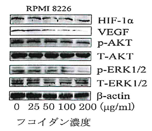
(Fucoidan concentration)
Fig.1)Effect of fucoidan on the expression of angiogenesis-related factors
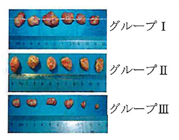
(Group I, Group II, Group III)
Fig.2)Suppressive effect of fucoidan on myeloma tumor formation
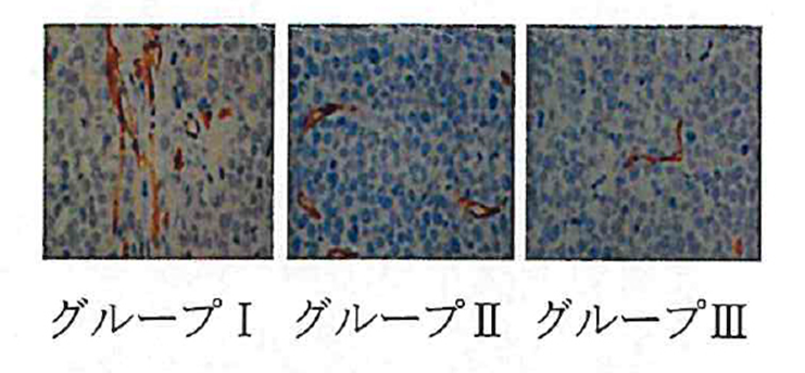
(Group I, Group II, Group III)
Fig.3)Angiogenesis-suppressing effect of fucoidan on myeloma